Our research interest is in bioanalytical chemistry.
Current effort in our group is directed towards spectroscopic imaging for cancer diagnosis and investigation of microscopic details in porous materials.
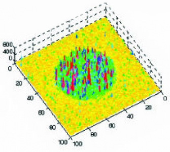
Early diagnosis is the key to improved treatment of cancers. If the cancer is diagnosed at an early stage without spread, the five-year survival rate of the patient is over 90%. If the cancer has spread to remote organs at the time of diagnosis, however, the survival rate decreases to below 10%. Optical spectroscopy provides a promising alternative or supplemental to histopathology, the standard method of cancer diagnosis in clinics. Optical methods are less invasive, inexpensive, and the diagnosis can be in situ, without time delay. To be a successful technique for cancer diagnosis, an optical method is required to provide a high contrast between normal/hyperplastic and adenomatous/adenocarcenomatous tissues. In addition, the measurement needs to be fast for clinical applications. We are developing spectrally-resolved fluorescence correlation imaging for cancer detection. In this novel imaging technique, each tissue location is investigated by two-dimensional fluorescence correlation spectroscopy (2D FCS). By combining both spectral and temporal information of the tissue fluorescence, 2D FCS provides a high contrast between samples, forming the basis for cancer diagnosis. In conjunction with tissue spectroscopy, fluorescence imaging and chemical separations are used to elucidate the molecular and morphological changes in tissue upon cancerous transformation.
Using imaging fluorescence correlation spectroscopy and single molecular spectroscopy, we are investigating interfacial behaviors inside nanometer sized pores. Our interest in porous materials is motivated by their usage as the stationary phases in chromatographic separations, as carriers for biomarkers, as vehicles for drug delivery and as support in catalysis. The material performance in all applications involves the diffusion, adsorption, and transport of molecules in the nanometer sized pores. These dynamic processes are largely influenced by the physiochemical properties of the materials that can be tuned with pore size, pore structure and surface modification. We are investigating these processes at the single molecule level. Studies are directed towards the adsorption and diffusion of molecules at the interface, the transport in confined dimension of nanometer sized pores, and the material microheterogeneity. Insights will help improve the technical performance of these materials in chemical and biological applications. For example, peak tailing and band broadening are two factors that degrade performance in chromatographic separations. Confocal imaging provides an excellent tool for investigating microscopic origins of band broadening and probing surface adsorption that is responsible for peak tailing.